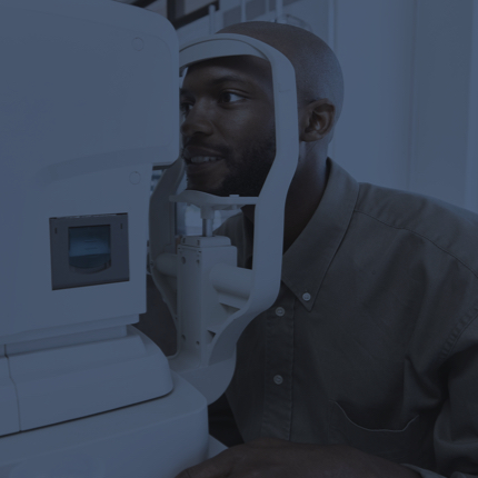
After lensometry and autorefraction are completed (see the Part 1 article last month), the pretest then focuses on ocular health screening. At some of our clinics, your intraocular pressure will be measured by the technician during pretest. This technique is called non-contact tonometry (NCT). You will be asked to remain still and keep your eyes open while a tiny puff of air is directed at the cornea. This may be slightly startling but our technician will prepare and reassure you. It takes only seconds for the intraocular pressure to be measured during this brief flattening of the cornea that does not require anaesthetic drops. If the results are normal and nearly equal between the eyes, this may be the only intraocular pressure measurement that is needed during your eye exam. If you have glaucoma or are a glaucoma suspect, or if the pressure is high or unequal between the eyes, your Optometrist will re-check the intraocular pressure in the exam room using eye drops and a different technique. Every full adult exam will include an intraocular pressure measurement, as this is important to screen for and manage ocular diseases like glaucoma.
The next pretest step is an automated visual field screening. One eye will be tested at a time. The patient is given a response button and is positioned so they are comfortably looking inside the instrument. They are asked to focus on a target at the center of the screen while pressing the response button each time they see a stimulus (generally a flickering grid pattern) in their peripheral vision. This stimulus is presented at various locations and intensities. The screening test generally takes less than one minute per eye. If a visual field defect is detected, the optometrist can then decide if further, more rigorous visual field testing is required. Visual field loss can occur with many conditions including glaucoma, stroke, and brain tumours.
The final portion of the pretest involves obtaining scans of the optic nerve and macula and images of the retina. Optical Coherence Tomography (OCT) is a non-invasive imaging technique that uses light waves to capture highly detailed scans of the retina. The thickness of the nerve fiber layer surrounding the optic nerve is measured. This provides important information regarding a patient’s risk of glaucoma and is also used to monitor changes in patients diagnosed with glaucoma. Scans of the macula show details of all the retinal layers and are important in conditions such as age-related macular degeneration, diabetic macular edema, and many other pathologies. Widefield retinal photos are captured with the Optomap camera. These images can show many ocular health conditions including retinal detachment, retinal vein occlusions, hypertensive retinopathy, and intraocular tumours. The detail of these scans and images is incredible and your Optometrist will be happy to show you the results as part of your eye exam experience.